Uni-Logo
Document Actions
  • We apply various configurations of the patch-clamp technique (whole-cell, cell-attached, excised inside-out, outside-out) to native and genetically manipulated cells and subcellular compartments. They enable us to monitor protein function and protein-protein interactions at high-resolution.
  • We use a large spectrum of biochemical techniques to detect and quantify membrane proteins (mainly ion channels and receptors), their post-translational modifications and association with other proteins in complexes and protein networks.
  • Modern mass spectrometers coupled with liquid chromatography enable us to identify several hundreds of proteins from complex samples with high confidence and sequence coverage. In addition, they provide quantitative data that let us determine stability, specificity and stoichiometry of protein-protein interactions as well as absolute protein abundance.
  • Nuclear magnetic resonance spectroscopy (NMR) provides information on structure and dynamics of biological macromolecules at atomic resolution under near-physiological conditions. We use it to examine proteins participating in the nano-environment of membrane proteins with regard to their 3D structure, mobility and interactions.
  • Using innovative microsystems, we work to enhance resolution and throughput of electrical recording of ionic currents. We develop biohybrid sensing devices based on single biological nanopores in membrane microarrays and study the interaction of natural and synthetic polymers with pore-forming membrane proteins.
  • To understand how neurons collectively process information, we develop optogenetic tools as well as new technologies for recordings from neurons in vivo and imaging of cell activity using photon Ca2+ and functional approaches. With computational network models we gain information on the principles underlying information processing in complex neuronal circuits.

Nuclear Magnetic Resonance Spectroscopy (NMR)

KChIP Structure (large)
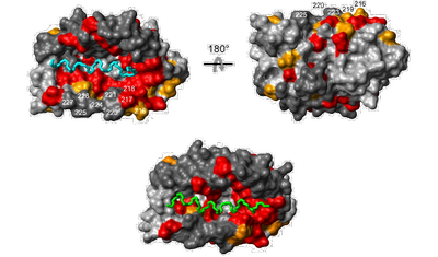
The α-helices of the KIS domain (green) and the Kv4.3 N terminus (cyan) can bind to the same surface pocket on the KChIP core structure as shown by mapping of NMR chemical shift perturbation data onto a surface representation of KChIP.

Practically, NMR studies of proteins imply

  • the expression and purification (mg amounts) of suitably isotope-labeled (15N, 13C, 2H) proteins for heteronuclear NMR experiments and
  • the recording and processing of NMR spectra (1D, 2D, 3D, …) followed by
  • a sequence-specific resonance assignment (protein backbone and side chains) or other types of spectral analysis.

Our institute is equipped with a Bruker Avance 600 NMR spectrometer with a cryoprobe allowing the investigation of proteins up to a size of 30-40 kDa at relatively low sample concentrations (< 300 µM).

3D structure determination by NMR requires NOE assignment and the measurement of additional structural constraints such as J coupling constants, residual dipolar couplings and paramagnetic relaxation enhancement (PRE). These data are then used as input for structure calculations which result in a family of structures.

Sequence-specific assignment and secondary structure of the extracellular domain of GLIC
Sequence-specific assignment and secondary structure of the extracellular domain of GLIC (yellow: assigned, light blue: unassigned, dark blue: proline).

Recent examples of our work are

  • the analysis of a potassium channel-interacting protein (KChIP) that revealed the structural basis for control of surface expression of Kv4 channel complexes by an autoinhibitory domain in the KChIP4a subunit [Schwenk et al., J Biol Chem., 2008].
  • the determination of the structural and dynamical properties of the monomeric extracellular domain of a prokaryotic nAChR homologue from the bacterium Gloeobacter violaceus [Chasapis et al., Biochemistry, 2011].
Personal tools